MRA Imaging: What It Is and How It Helps Diagnose Vascular Conditions
When doctors need to see your blood vessels without cutting you open, they often turn to MRA imaging, a type of magnetic resonance imaging that focuses specifically on blood vessels to detect blockages, aneurysms, or abnormalities. Also known as magnetic resonance angiography, it uses powerful magnets and radio waves to create detailed pictures of arteries and veins—no X-rays needed. Unlike traditional angiograms that require catheters and contrast dyes injected into arteries, MRA is non-invasive and safer for most people, especially those with kidney issues or allergies to iodine-based contrast.
MRA imaging is closely tied to vascular imaging, the broader field of techniques used to visualize blood flow and vessel structure. It’s one of the most accurate tools for spotting problems like carotid artery stenosis, pulmonary embolisms, or aortic dissections. It also plays a big role in evaluating brain circulation, especially after a stroke or transient ischemic attack. Doctors use MRA to check for aneurysms in the brain before they rupture, or to see if a clot is blocking blood flow to the heart or limbs. The images it produces are so detailed that surgeons often rely on them to plan operations—like removing a tumor near a major artery or placing a stent.
What makes MRA stand out is how it avoids radiation. While CT angiograms give fast results, they expose you to ionizing radiation. MRA doesn’t. That’s why it’s often preferred for younger patients, pregnant women, or anyone needing repeated scans over time. It’s not perfect—some people with metal implants can’t have it, and claustrophobia can be an issue. But for most, it’s the best balance of safety and clarity.
Behind every MRA scan is a team that knows how to interpret the data. Radiologists look for subtle changes in blood flow patterns, vessel wall thickness, or abnormal bulges that might mean trouble. These aren’t just pictures—they’re clues to what’s happening inside your body. And with advances in software, newer MRA machines can even show real-time blood movement, helping doctors predict where a clot might travel next.
Below, you’ll find real-world insights from patients and providers who’ve used MRA imaging to understand their conditions better. From how it helped spot a hidden aneurysm to why some people get confused by the results, these posts cut through the noise and give you the facts you need—no jargon, no fluff, just what matters.
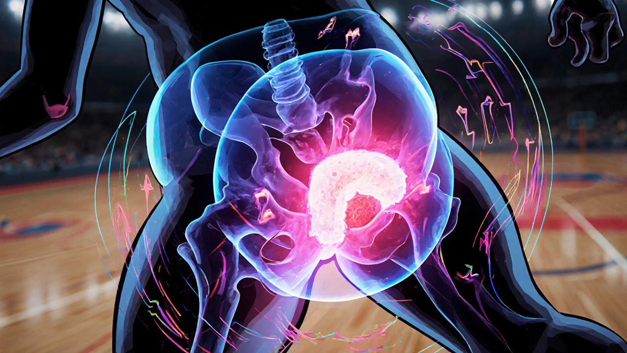
Hip Labral Tears in Athletes: Diagnosis, Imaging, and Arthroscopy Recovery
Nov, 24 2025
Hip labral tears are common in athletes and require accurate diagnosis with MRA imaging. Treatment ranges from physical therapy to arthroscopic repair, with recovery taking 3-6 months. Addressing underlying bone issues is critical to prevent re-tears and early arthritis.